
On the Ear.
The Human Ear is separable into three distinct parts - the External ear, the Middle ear, and the Internal ear.
The External ear consists of the cartilaginous lobe or Auricle, and the External Meatus (E. M. fig. 16). The latter is a tube about an inch and a quarter long, directed inwards and slightly forwards, and closed at its inner extremity by the Tympanum (Ty., fig. 16) or drum of the ear, which is stretched obliquely across it.
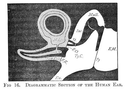
The Middle ear, which is separated from the External ear by the Tympanum is a cavity in the bony wall of the skull, called the Tympanic Cavity (Ty. C., fig. 16). From this, a tube, about an inch and a half long, termed the Eustachian Tube (Eu., fig. 16) leads to the upper part of the throat, and thus places the air in the Tympanic Cavity in communication with the external air. On the side opposite to the tympanum, there are two small appertures in the bony wall of the cavity, both of which, however, are closed with membrane. The lower of these two apertures, which is about the size of a large pin's head, is called from its shape the Fenestra Rotunda, or round window (F.R., fig. 16); the upper and rather larger one, the Fenestra Ovalis, or oval window (F.O., fig. 16).
A chain of three small bones, stretches between the Tympanum and the Fenestra Ovalis. One of these, the Malleus or Hammer Bone (Mall., fig. 16) is firmly fastened by one of its processes or arms to the Tympanum, while the other process projects upwards into the cavity, and is articulated to the second bone - the Incus or Anvil (Inc., fig. 16). The lower part of this last again, is articulated to the third bone - the Stapes or Stirrup Bone (St., fig. 16), which in its turn is firmly fastened by the flat end of the Stirrup Bone to the membrane which closes the Fenestra Ovalis. These three bones are suspended in the tympanic cavity in such a way, that they are capable of turning as a whole upon a horizontal axis, which is formed by processes of the Incus and Malleus, which processes fit into depressions in the side walls of the cavity. This axis in fig. 16 is perpendicular to the plane of the paper, and would pass through the head or upper part of the Malleus.
There are also two small muscles in the tympanic cavity, by the contractions and relaxations of which, the membrane of the Fenestra Ovalis and the tympanic membrane can be rendered more or less tense. One, called the Stapedius, passes from the floor of the tympanic cavity to the Stapes, and the other - the Tensor Tympani - from the wall of the Eustachian tube to the tympanum.
Before going further, let us consider the functions of these various parts. The sound waves which enter the External Meatus, pass down it, assisted in their passage by its configuration, and strike against the Tympanum. Now, as the air in the cavity of the tympanum is in communication with the outer air, as long as the latter is at rest, they will be of the same density, and hence the Tympanum will sustain an exactly equal pressure from both sides. When, however, the condensed part of a sound wave comes into contact with it, this equilibrium will no longer exist; the air on the outer side will exert a greater pressure than that on the inner, and the Tympanum will bulge inwards in consequence. The condensed part of the wave will be immediately followed by the rarefied part, and now the state of things is reversed; there will be a greater pressure from the inside, and consequently the Tympanum will move outward, a movement assisted by its own elasticity. We see therefore, that the Tympanum will execute one complete vibration for every wave that reaches it; that is, as we know from the preceding chapter, it will perform one vibration for every vibration of the sound-producing body.
Further, it follows from the arrangement of the Malleus, Incus, and Stapes, and the attachment of the first and last to the Tympanum and the Fenestra Ovalis respectively, that every movement of the Tympanum must cause a similar movement of the Fenestra Ovalis. We see, therefore, that this latter membrane faithfully repeats every vibration of the Tympanum, that is, every movement of the sound-originating body.
The Internal ear is of a much more complicated nature than the parts already described. It consists essentially of a closed membranous bag of a very irregular and intricate shape, which contains a liquid termed Endolymph, and which floats in another liquid called Perilymph. Both the Endolymph and the Perilymph are little else than water. This membranous bag may be divided, for the purpose of explanation, into two parts - the Membranous Labyrinth (M.L., fig. 16) and the Scale Media of the Cochlea (Sca. M., fig. 16).
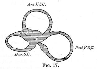
The Membranous Labyrinth comprises the Utriculus (Ut., fig. 17, the Sacculus, and the three Semi-circular Canals. All these parts communicate with one another, forming one vessel; the two first lying one behind the other, and the three canals springing from the Utriculus. One end of each of the Semi-circular Canals is dilated at the point where it joins the Utriculus into a swelling called an Ampulla (A, fig. 17). Of the three canals two are placed vertically and the other horizontally; hence their names - the Anterior Vertical Semi-circular Canal (Ant. V.S.C., fig. 17), the Posterior Vertical Canal (Post. V.S.C., fig. 17) and the Horizontal Canal (Hor. S.C., fig. 17). All these parts are contained in a bony casing, which follows their outline pretty closely, so that there is a bony labyrinth, bony semi-circular canals, and so on. There is however a space between the bony casing and the membranous parts, which is filled by the Perilymph referred to above.
Every parts of the Membranous Labyrinth is lined by an exceedingly delicate coat - the Epithelium - the cells of which, at certain parts, are prolonged into very minute hairs, which thus project into the Endolymph. In close communication with these cells are the ultimate fibres of one branch of the Auditory Nerve, which, ramifying in the wall of the Membranous Labyrinth, pierces its bony cassing and proceeds to the brain. Floating in the Endolymph, there are also minute hard solid particles called Otoliths.
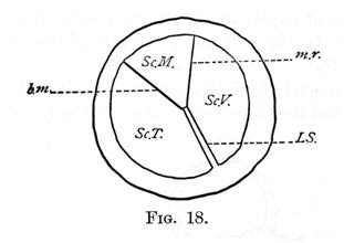
The remaining part of the Internal ear is more difficult to describe. It consists essentially of a long tube of bone closed at one end, of which fig. 18 is a diagrammatic section. A thin bony partition - the Lamine Spiralis, L. S. - projecting more than half way into the interior, runs along the tube from the bottom, nearly but not quite to the top, so that the two chambers - Sc. V. and Sc. T - communicate at the closed end of the tube. These two chambers - the Scala Vestibuli and the Scala Tympani - are filled with Perilymph. Diverging from the interior edge of the Lamina Spiralis and terminating in the bony wall of the tube, are two membranous partitions, the Membrane of Reissner - m.r. - and the Basilar Membrane - b.m. The chamber between these two membranes which is termed the Scala Media - Sc.M. - is filled with Endolymph. So far the tube has been described for simplicity as if it were straight. Now, we must imagine it forming a close coil of two and a half turns round a central axis of bone - the Modiolus; the Scala Media being outwards and the Lamina Spiralis springing from the axis. The whole arrangement is not unlike a small snail's shell and is termed the Cochlea. It is so placed with reference to the other parts of the ear, that the Scala Tympani is closed at the lower end by the Fenestra Rotunda (F.R., fig. 16) while the Perilymph of the Scala Vestibuli is continuous with that of the Labyrinth. On the other hand, the Endolymph which fills the Scala Media is in communication with that of the Sacculus.
Resting on the upper side of the elastic Basilar membranes are the arches or rods of Corti. Each of these rods consists of two filaments, joined at an angle like the rafters of a house. Altogether there are some three or four thousand of them, lying side by side, stretching along the whole length of the Scala Media like the keys of a pianoforte. A branch of the auditory nerve, entering the Modiolus, gives off fibres which pass through the Lamina Spiralis, their ultimate ramifications probably coming into close connection with Corti's rods.
Not much is known for certain of the functions of the various parts of the Internal ear. We have seen how the vibrations of the sounding body are transmitted to, and imitated by, the membrane of the Fenestra Ovalis. These vibrations are necessarily taken up by the Perilymph, which bathes its inner surface, and hence communicated to the Endolymph of the Membranous Labyrinth and the Scala Media. It was formerly thought, that both of these organs were necessary to the perception of sound, but the researches of Goltz have shown that the special function of the Labyrinth, is to enable us to perceive the turning of the head. Thus, the only part of the Internal Ear which is engaged in transmitting sound vibrations to the auditory nerves, is the Cochlea.
The Basilar Membrane of the Cochlea consists of a series of radial fibres, lying side by side, united by a delicate membrane, and it is believed by Helmholtz, that the faculty of discriminating between sounds of different pitch is due to these radial fibres. The Basilar Membrane gradually increases in width, that is, its radial fibres gradually increase in length, as we pass from the Fenestra Ovalis to the apex of the Cochlea, being ten times as long at the latter, as at the former. Helmholtz likens them to a series of stretched strings of gradually increasing lengths, the membranous connection between them simply serving to give a fulcrum to the pressure of the fluid against the strings. He further assumes that each of these fibres is tuned to a note of definite pitch, and capable of taking up its vibratory motion. He considers that the arches of Corti which rest on these fibres, serve the purpose of transmitting this vibratory motion to the terminal appendages of the nerve, each arch being connected with its own nerve ending.
As already mentioned, there are about 3,000 of these arches in the human ear, which would give about 400 to the octave. When a simple tone is sounded in the neighbourhood of the ear, the radial fibre of the Basilar Membrane in unison with it, is supposed to take up its vibrations, which are then transmitted by the arch of Corti in connection with it, to the particular nerve termination with which it is in communication.
For a complete discussion of this theory, the reader is referred to Helmholtz's "Sensations of Tone," Part I, Chap. vi.
Summary.
The Human Ear may be divided, for descriptive purposes, into three parts: the External ear, Middle ear, and Internal ears.
The External ear consists of the Lobe, and the tube or Meatus leading inwards, which is closed by the Tympanum.
The Middle ear contains a chain of three small bones, the Malleus, Incus, and Stapes, which serves to connect the Tympanum with the Fenestra Ovalis. The air in the cavity of the Middle ear is in communication with the external air, by means of the Eustachian Tube.
The Internal ear consists essentially of a membranous bag of exceedingly complicated form, filled with a liquid - Endolymph. This bag floats in another liquid - Perilymph - contained in a bony cavity, which is separated from the cavity of the Middle ear by the membranes of the Fenestra Ovalis and Fenestra Rotunda.
The nerves of hearing ramify on the walls of this membranous bag, and their ultimate fibres project into the Endolymph therein contained.
The alternate condensations and rarefactions of the sound waves which enter the External ear, strike against the Tympanum and set it vibrating. These vibrations are then transmitted by means of the small bones of the Middle ear to the Fenestra Ovalis, and from this again through the Perylimph and Endolymph to the minute terminations of the auditory nerve, which lie in the latter liquid.
One particular part of the membrane of the Internal ear, in which lie the Fibres of Corti, is specially modified, and is supposed to be the region of the ear which serves to discriminate the pitch and quality of musical sounds.
Ramsay
Effect here means, of course, musical effect in the ear's appreciation of the notes. [Scientific Basis and Build of Music, page 28]
See Also